Fracture Repair
When your pet has a broken bone and you’re searching for the right veterinary hospital to perform the surgery, there are some important considerations to consider:
Experience: At our office, orthopedic surgery is something we do on a daily basis. We perform approximately 600 orthopedic surgeries each year, making us one of the most experienced clinics in the bay area.
Cost: Our total cost on most advanced procedures tends to be about half that of the larger, corporate specialty centers. We do all of our orthopedic surgeries with the same level of care and quality. Given that our volume of these procedures is much higher than other most places, we are able to keep costs lower for each patient.
Availability: We have multiple doctors who exclusively perform orthopedic surgery at our office and we put time aside in our schedules to accommodate more urgent procedures - such as fracture repairs. With fractures, time is of the essence. Irreversible damage may be done when there is a 1 or 2 week delay between the injury and surgery date.
Reputation: We’ve been lucky to be one of the highest-rated clinics in Alameda County. Many clients come to us through word-of-mouth and also dozens of area veterinary clinics have referred their patients to us for orthopedic surgery.
There are a lot of factors to consider when we decide how best to fix a broken bone. A few of these factors include: How old is the patient? How old is the fracture? Which bone is broken? Did the bone break through the skin? Are there multiple bone fragments involved? Is the pet calm or will they be overactive after the surgery?
Ilium Fracture
This was a younger, large dog with a broken left Ilium, which is part of the hip. You can see that the left and ride sides of the hip are no longer symmetric and one joint is above the other one. We repaired this fracture with a bone plate which needed to be contoured to the curve of the hip bone. Hip fractures are typically more difficult than other fractures due to the curve of the bone and its location deep within the muscles. This patient healed well within 2 months after surgery.


Femur Fracture
This fracture occurred in a younger, indoor-outdoor, cat. We repaired it by a method called Rush Pinning. This method was chosen because the broken distal fragment of bone was too small to accept a bone plate.


Tibia Fracture
This fracture occurred in a younger cat. What makes a case like this worse is that it’s a comminuted fracture - meaning that there are multiple fragments rather than one clean break. This has to be taken into account when deciding on how best to fix the leg. We contoured and placed a small bone plate along the inner surface of the tibia to hold the leg in place as the bone healed.


Femur Fracture
This fracture occurred in a middle-aged, toy breed dog. Because the fracture happened closer to the middle of the bone, in contrast to the x-rays from above, we were able to place a very small bone plate against the outer surface - which provided the best stability.


Examples from our office
Humerus Fracture
This surgery was done in a younger cat. Humerus fractures can be more difficult than other bones due to the curved shape of the bone and need for extra stability. We repaired this fracture using both an IM pin and a lateral locking bone plate and screws. Sometimes this combined approach is best to give both bending and rotational stability.


Humeral Condylar Fracture
This injury happened in a 7 month old toy breed dog who fell off the couch and landed wrong. This type of fracture is common in French Bulldogs and other toy breeds. It can happen at any age but is more common in younger pets. In this case, after “reducing” (putting back together) the fracture, it was held in place with a lag screw to compress and stabilize the fracture line and a k-wire for anti-rotation support.

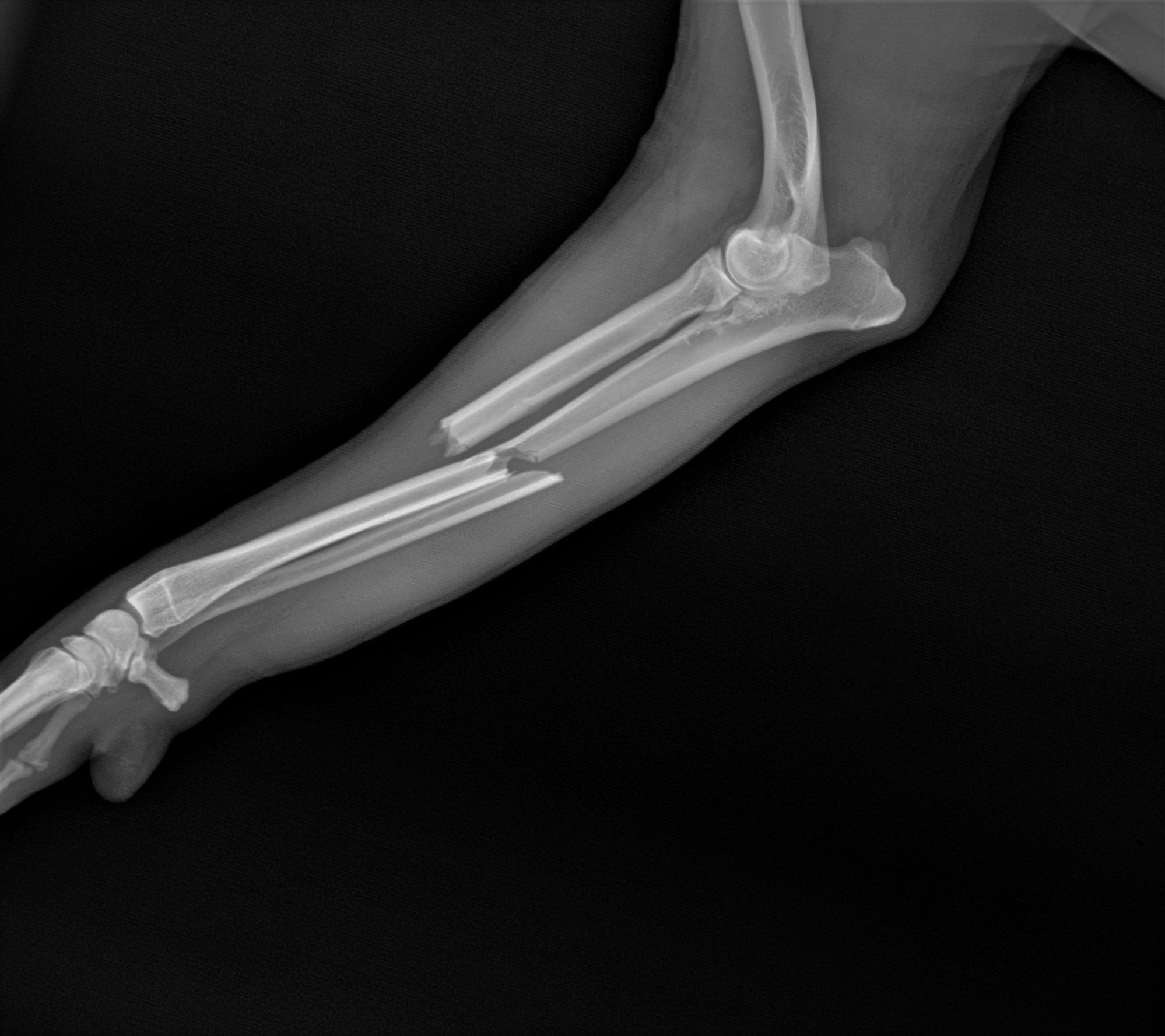
Radius/Ulna Fracture
This surgery was done in a very small toy breed dog (7lbs). Toy breed pets can be especially difficult because the size of the implant needed is exceedingly small. The screws used in this case were just 1.5mm in diameter.


Tibial Tuberosity Avulsion
In young dogs, the growth plates are still open and prone to fracture. Sometimes, along with a traumatic event, the patellar tendon pulls part of the tibia right off the front of the bone. To properly secure this back in place, we placed 2 thin wires - called Kirschner Wires. In this particular case, the after photo is from 8 weeks later, showing how the bone has fully healed.


Ununited Anconeal Process
This was a younger, larger, German Shepherd with a condition called elbow dysplasia. In this disease, portions of the elbow don’t develop properly, and this can be painful. In this dog, the anconeal process was separated from the rest of the ulna. We put it back in place, held there with a wire and surgical headless compression screw. In addition, we performed a dynamic ulna osteotomy (DUO), which helps restore joint congruity as the anconeal process heals.


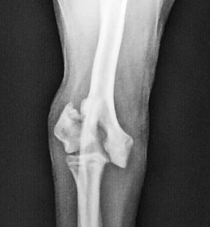
Radius/Ulna Fracture
This surgery was performed on a medium sized dog. The difference from the above fracture is that this break occurred in the middle of the bones - not on the end. Therefore, a slightly different approach was used.